

Anatomically correct –
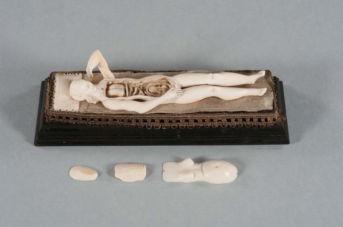
The carved figurines measure 4 to 9 inches long and include tiny removable organs.
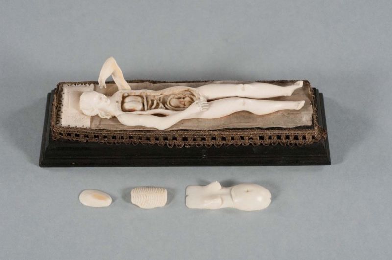
Enlarge/An ivory manikin after removal of the abdominal and chest wall, ribs, and part of the uterus. Internal organs such as the lungs, intestines, as well as a fetus inside the uterus are visible.
Researchers at Duke University have completed digital scans of small medical manikins and identified the materials used to make them. They will be presenting their findings next week at the annual meeting of theRadiological Society of North America(RSNA) in Chicago.
These are not the department-store mannequins familiar to most of us today. Rather, they are tiny, intricately carvedanatomical figurinesdating back centuries.According to the New York Academy of Medicine, there was an explosion of interest in three-dimensional anatomical models in the mid – 16 th century, typically made of wax molds or carved from wood or ivory. The manikins likely emerged as a result of this trend. Scholars have pegged their origins to Germany in the late 1600 s or early 1700 s, possibly created in the Nuremberg workshop of sculptor Stephan Zick, known for his ivory models of human ears and eyeballs.
The figurines measure between 12 to 24 centimeters (4 to 9 inches) and have movable arms. The torso has a lid that can be removed to reveal tiny, intricately carved organs within the cavity. The removable organs include lungs, heart, intestines, bladder, kidneys, stomach, liver, and pancreas. There are some male and female pairs, but most of the manikins are pregnant female figures, with a tiny carved fetus attached to the uterus with a red cord.
What are they good for?
Exactly what the manikins were used foris unclear. Historians don’t believe they were teaching aids, given the lack of intricate details of the carved internal organs. It’s been suggested they may have been used by doctors to explain childbirth to their pregnant female patients, butart historian Cali Buckley, among others, thinks it is far more likely they were collectibles, popular with male physicians as a display of luxury or wealth.
Duke University has one of the largest collections of the surviving manikins: 22 of the 180 known to be in collections around the world. The figurines were donated in 1956 from the private collection of a thoracic surgeon at Duke named Josiah Trent. They are fragile items, susceptible to light damage, and thus are rarely displayed. “They are usually stored in a library vault and occasionally rotated into a special display unit in the Duke Medical Library for visitors to appreciate,”said co-author Fides R. Schwartz, a research fellow in the Department of Radiology at Duke.
Two years ago, university conservationists teamed up with the Shared Materials Instrumentation Facility (SMiF) to use micro-CT scanningto digitizethe entire collection. The objective was to preserve the fragile pieces in digital form, as well as enabling 3D printing of replicas for more frequent display.
-
Male and female manikins from the collection in the Wellcome Institute of the History of Medicine.
-
Male and female manikins from the collection in the Wellcome Institute of the History of Medicine.
-
Duke University’s 3-D X-ray scans reveal how the manikins were put together or repaired as they changed hands over the centuries.


GIPHY App Key not set. Please check settings