

Under the microscope –
The Olympus Global Image of the Year honorees find beauty under the microscope.
As Ars’ John Timmer noted
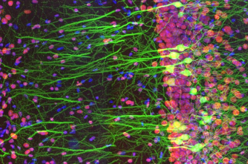
Spain’s Ainara Pintor snagged the top honor from over 793 submissions with her gorgeous image of an immunostained mouse-brain slice, titled Neurogarden . The image focuses on the hippocampus area of a single slice, but there are more than million neurons in the mouse brain as a whole, according to Pintor. Howard Vindin of Australia won the regional prize for Asia-Pacific by capturing an autofluorescence image of a mouse embryo. US entrant Tagide de Carvalho won the regional award for the Americas with his colorful image of a tardigrade. The regional winner for Europe, the Middle East, and Africa was the UK’s Alan Prescott, for his image capturing the frozen section of a mouse’s head.
Honorable mentions included striking microscopic images of photonic crystals in insect scales, crystallized amino acids, desert locust wings, and opal embedded in iron sandstone, among others. Clearly, the field of photomicroscopy is still attracting top-notch talent.
Global Winner: immunostaining of a mouse brain slice with two fluorophores
Regional Winner for the Americas: it’s a colorful tardigrade!
Tagide de Carvalho / Olympus
Asia-Pacific Regional Winner: Autofluorescence image of a mouse embryo.
Howard Vindin / Olympus
EMEA Regional Winner: frozen section of a mouse’s head.
Alan Prescott / Olympus
Honorable Mention: mouse spinal cord.
Tong Zhang / Olympus
Honorable Mention: image of 3D depth color-coded reconstruction of confocal images of microtubules in monkey fibroblast cells.
Daniela Malide / Olympus
Photonic crystals in insects (beetles and weevils) FTW!
Rudolf Buechi / Olympus
Preparation of amino acids crystallized out of an ethanol solution.
Justin Zoll / Olympus
The desert locust is one example of an insect species that has evolved foldable wings, the better to keep them clean and intact. This image is called A Road in the Sky because “veins look like roads and spines on [the] wing membrane are like stars.”
Hamed Rajabi / Olympus
Green gem material, prase opal, magnified through a microscope, looks like an aerial coastline view. The brown areas are iron sandstone host rock.
Nathan Renfro / Olympus
Microscopic image of fruit fly brain.
Martin Hailstone / Olympus
Inflorescence of developing flower buds expressing fluorescent reporters (cyan), with cell walls stained red.
Nat Prunet / Olympus
The ovary of a gall-inducing wasp showing the eggs.
Ming-Der Lin / Olympus

GIPHY App Key not set. Please check settings